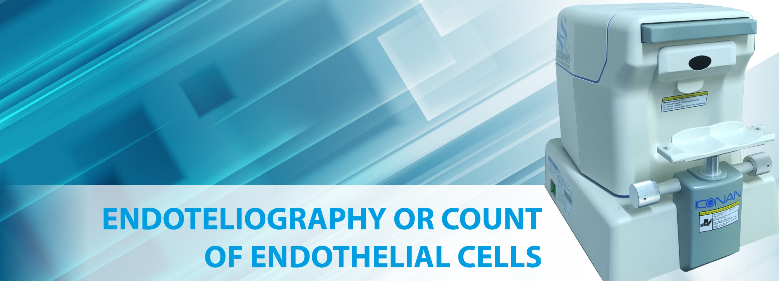
This examination is carried out with a Specular Microscope, equipment that allows one or several photographs to be taken of the innermost layer of the cornea. Once the image is obtained, the technician must count the number of cells found within a certain field in square millimeters and the ophthalmologist can evaluate the quality of the tissue in terms of the number, shape and size of the endothelial cells.
With this examination, the treating ophthalmologist has the possibility of diagnosing corneal diseases and dystrophies, pre- and post-surgical follow-up of cataract surgery, corneal transplant or keratoplasty, corneal decompensation, suspicion of dystrophies, trauma, etc.
Horarios de toma de examen:
Monday to Friday from 7:30 a.m. to 12:30 p.m. and from 2:00 p.m. to 4:00 p.m.
Delivery of results:
Results will be available 3-5 business days after the exam is taken.
You can request them in person from Monday to Friday 7:30 a.m. to 5:00, Saturdays 7:30 a.m. 12:30 pm. p.m. Or by email archivo@barraquer.com.co


