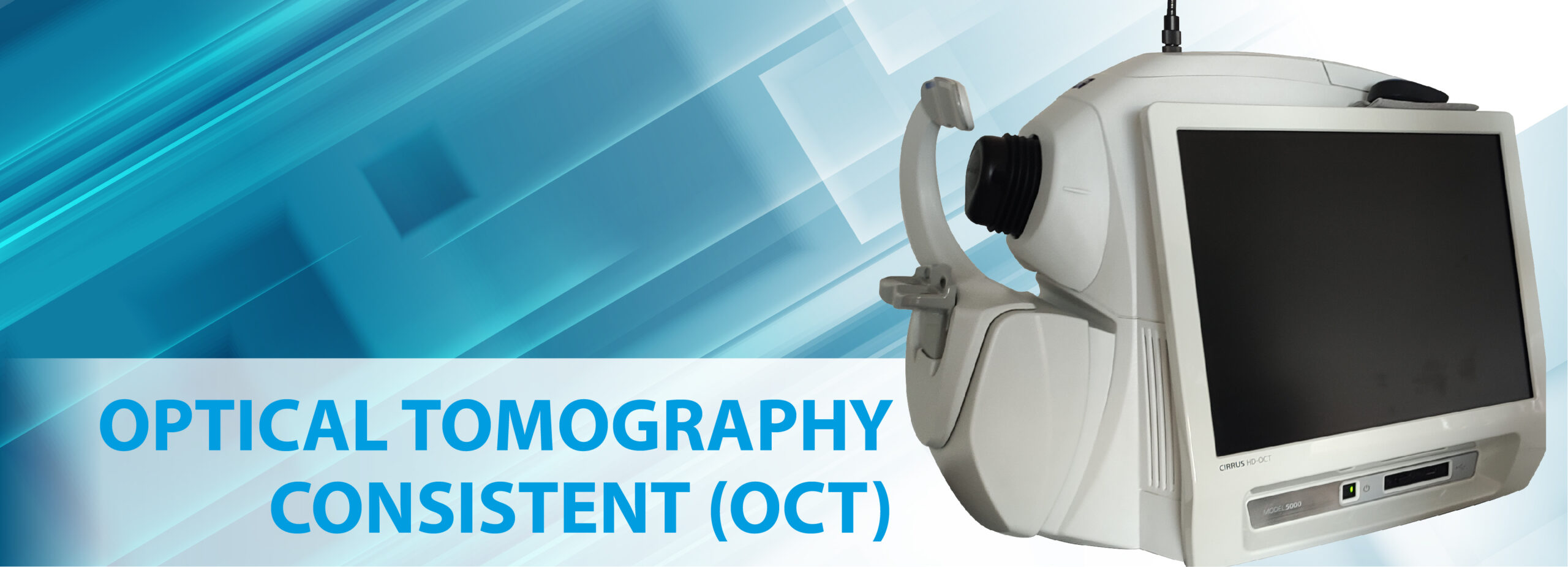
The OCT or Optical Coherence Tomography is a diagnostic test that detects changes in the entire retinal structure, covering each of the 10 layers that compose it, specifically in the macula (central part of the retina) and the Optic Nerve, in the segment posterior part of the eye and in the anterior part of the eyeball in Cornea and the iridocorneal Angles or Camerular Angles.
In general, it is an exam that is carried out from the taking of several scans with different functions that the equipment has and through the images obtained, pathologies in the retina and optic nerve can be diagnosed.
OPTICAL COHERENT TOMOGRAPHY CLASSIFICATION – OCT
THE MACULA OCT. It is a non-invasive examination of the central part of the retina that allows diagnosing, monitoring pre- and post-surgical treatments and controlling diseases of this tissue. Macular Tomography is extremely useful in the diagnosis of retinal diseases such as age-related macular degeneration (AMD), macular edema, macular hole, diabetic retinopathy, central serous choroidopathy, hypertensive retinopathy, among many others.
In general, the risk factors for these diseases are usually age, genetic inheritance (diabetes, hypertension, retinitis pigmentosa). Others may be the result of some type of trauma or may also occur spontaneously. In any case, these pathologies can seriously affect visual capacity. That is why it is important to attend the ophthalmologist regularly for early detection of pathologies, prevention and control of them, all the more so if the patient is over 50 years of age.
OPTIC NERVE OCT. It is a non-invasive examination of the Optic Nerve, which through scanner-type images allows an analysis of the nerve fiber layer (NFL) that is around the optic disc, the cup/disc relationship with high resolution and the analysis of ganglion cells. to detect early the disease that occupies the first place of cause of blindness in the world: Glaucoma. It also plays a very important role in monitoring patients with this disease, allowing comparative charts to be made over the years. It is also indicated for other types of optic nerve pathologies such as drusen, optic neuritis, papilledema or papilledema, etc.
CORNEA OCT. Test performed on the first lens of the eye called the cornea. Through images that show each of the 5 layers of the cornea, it is possible to diagnose, treat and control pathologies by analyzing the structure of this organ. Useful for the evaluation of different pathological conditions such as keratoconus, corneal ulcers, abscesses, corneal dystrophies, foreign bodies, bullous keratopathy, leukomas and corneal scars, as well as in the pre and postoperative study of refractive surgery using Excimer Laser (after this intervention the scar forms precisely on the cornea).
OCT OF CAMERULAR ANGLES. It is a very precise examination in which, through exact scan-type images, the measurement of the iridocorneal angles or chamber angles can be obtained, which is very useful for classifying open-angle and angle-closure glaucoma, providing the necessary information to make management decisions. specific to each.
Exam taking times:
Monday to Friday from 7:30 a.m. to 12:30 p.m. and from 2:00 p.m. to 4:00 p.m.
Delivery of results:
Results will be available 3-5 business days after the exam is taken.
You can request them in person from Monday to Friday 7:30 a.m. to 5:00, Saturdays 7:30 a.m. 12:30 pm. p.m. Or by email archivo@barraquer.com.co